VP image

NO SWIMMING EVER AFTER SURGERY! Image Courtesy Lori Shriver
Most of us know the larynx is commonly known as the voice box and is located in the throat. We know that laryngitis is a condition where one cannot speak, but other than that, the larynx does not get much thought. It is a vastly underappreciated organ. The larynx is not just where the sound comes from; it is, more importantly, the cap of the respiratory tract's tubing. The larynx closes the respiratory tract off while we eat and drink so that we do not inhale our food. If we need to take a deep breath, the muscles of the larynx expand and open for us. The larynx is the guardian of the airways, keeping whatever we want to swallow out and directing air in.
Laryngeal paralysis results when the abductor muscles of the larynx cannot work properly. This means no expanding and opening of the larynx for a deep breath; the laryngeal folds simply flop weakly and flaccidly. In other words, when you need a deep breath, you don't get one. This can create tremendous anxiety. imagine attempting to take a deep breath and finding that you simply cannot. Anxiety leads to more rapid, deep breathing attempts and more distress. A respiratory crisis from the partial obstruction can emerge, creating an emergency and even death.
Laryngeal paralysis does not come about suddenly. For most dogs, there is a fairly long history of panting, easily tiring on walks, or loud raspy breathing. Ideally, the diagnosis can be made before the condition progresses to an emergency.
Dogs with laryngeal paralysis demonstrate some or all of the following signs:
- Excess panting
- Exercise intolerance
- Voice change
- Raspy breathing sounds
- Respiratory gasping or distress
The usual patient is an older, large-breed dog; the most commonly affected breed is the Labrador retriever. The condition can occur in cats, but it is rare. The Bouvier des Flandres has a hereditary form of laryngeal paralysis that is able to affect young dogs. Other breeds reported to have early-onset genetic laryngeal paralysis include Siberian Husky, Great Pyrenees, bull terrier, and Dalmatian.
Is Laryngeal Paralysis Part of a Bigger Neurologic Problem?
The time for ambiguity regarding this question has passed, and the answer is a definite yes. Laryngeal paralysis is now considered to be the first symptom of a much more pervasive neurologic weakness, hence the new name of the condition, "Geriatric Onset Laryngeal Paralysis and Polyneuropathy." In time, the leg muscles will become weak and uncoordinated, leading to debilitating mobility problems. In addition, the esophagus (the tube that carries food from the throat to the stomach) will lose normal function, creating a risk of inhalation of food material. This can progress to a completely flaccid esophagus called a megaesophagus, a condition that requires high maintenance management to prevent aspiration pneumonia and provide proper food delivery.
The good news is that the average patient with acquired laryngeal paralysis is at least 10 years old, and the progression of the neurologic weakness is fairly slow. This means many patients will live their normal lifespan before further neurologic weakness becomes a problem. We can still say, however, that dogs with laryngeal paralysis are 21 times more likely to develop a megaesophagus than dogs without laryngeal paralysis. Also, if one knows that progressive muscle weakness is coming, one can be proactive with exercise and physical therapy to help maintain mobility as long as possible.
It's been suggested that hypothyroidism may be a cause of laryngeal paralysis. In fact, it is not. Hypothyroidism is associated with other neuropathies that could complicate the polyneuropathy of which laryngeal paralysis is a part. This means hypothyroidism should be identified and treated, but while improvement in weakness, etc., may be seen, the laryngeal paralysis will not reverse with thyroid hormone supplementation.
Making the Diagnosis
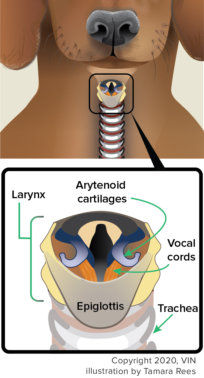
To determine if a dog has laryngeal paralysis, the larynx must be examined under sedation. The level of sedation must be heavy enough to allow the larynx to be inspected but light enough for the patient to take some deep breaths. If the sedation is too deep for the diagnosis to be obvious, a respiratory stimulant called Dopram® (doxapram hydrochloride) is given intravenously. This is to stimulate several deep breaths so that the function of the larynx is clear. In a normal larynx, the arytenoid cartilages are seen to open and close widely. In a paralyzed larynx, they just sit there limply while the patient breathes deeply.
If the patient is having a respiratory crisis when seeing the veterinarian, this diagnostic test can easily be followed by intubation, inserting a breathing tube down the patient’s throat. This relieves the upper airway obstruction, and the patient can breathe normally. Unfortunately, sedation must be maintained to keep the tube in place. This potentially poses a problem when the patient must wake up, and the tube must be eventually removed. If waking up involves too much respiratory motion, the patient may have to be re-sedated and the tube put back in until another attempt at waking can be managed.
A newer technique of visualizing the larynx involves threading an endoscope down the patient’s nostril. This is tricky, but the benefit is that sedation is not required. The downside is that specialized equipment is needed, and the patient may not be cooperative.
Additional Testing
There are some additional tests that are helpful in evaluating the patient with laryngeal paralysis. Chest radiographs are important in ruling out aspiration pneumonia (from inhaling food material through the non-functional larynx), megaesophagus (which we have mentioned tremendously complicates a laryngeal paralysis case), and obvious tumor spread. Radiographs of the throat to rule out obvious throat tumors are also helpful. Complete blood testing, including thyroid tests, should also be included in the workup.
Conservative Treatment
Treatment of laryngeal paralysis is most likely going to require surgery, but not everyone is ready or able to provide a surgical solution for their dog. Here are some tips for non-surgical management:
- Change from collar to harness to avoid pressure on the larynx.
- Avoid heat or other situations where the dog might pant.
- Reduce activity (also to reduce panting)
- Tranquilizers or anti-anxiety medications may have benefits.
Canine Larynx Diagram

The Crisis
If laryngeal paralysis is not treated, a respiratory crisis can emerge. In this situation, the patient attempts to breathe deeply and simply cannot, creating a vicious cycle of anxiety and respiratory attempts. The laryngeal folds become swollen, making the throat obstruction still worse. The patient’s gums become bluish from lack of oxygen, and the patient begins to overheat. For reasons that remain unclear, fluid begins to flood the lungs, and the patient begins to drown (as if the laryngeal obstruction wasn’t lethal enough).
The patient must be sedated, intubated, and cooled down with water to survive. As soon as intubation is in place, the patient can breathe normally, oxygen can be given, and the crisis can be curtailed if it has not progressed too far.
Of course, eventually, the patient will have to wake up and be able to survive without medical equipment. Corticosteroids can be used to reduce the swelling, but ideally, one of several surgical solutions is needed.
Surgical Solutions
The goal of surgery, whichever technique is used, is to relieve the airway obstruction permanently while maintaining the original function of the larynx (protection of the airways).
Laryngeal Tieback (also called Lateralization Surgery)
This has probably become the most commonly performed surgery for laryngeal paralysis. It involves placing a couple of sutures in such a way as to pull one of the arytenoid cartilages backward. By repositioning one of the arytenoids, the opening of the larynx is made larger. The chief complication is that only a few millimeters of position change in the arytenoids are needed. If the cartilage is moved too much, the larynx cannot properly close, and aspiration pneumonia becomes a substantial risk. Commonly, these patients have a persistent cough after eating or drinking. This surgery has been associated with a 14 percent postoperative mortality rate. (Years ago, both arytenoids were tied back to create a still larger larynx, but tying off both cartilages in this way was associated with a 67% mortality rate, so it is no longer done.)
Partial Arytenoidectomy
Another surgical technique involves only biting out one vocal fold and also biting out the arytenoid cartilage on the same side. There is more bleeding with this technique, and a tracheostomy becomes more desirable. Surgeries involving removing part of the larynx have been associated with a 30% mortality rate in laryngeal paralysis patients.
Ventriculocordectomy (De-Barking)
De-barking surgery is generally thought of as a surgical solution to a behavioral problem, but it is also a fair treatment for laryngeal paralysis. The usual method involves extending a long “biting” forceps down the throat and biting out the vocal folds. Anesthesia is required to do this, and the fact that the surgical area is the larynx makes normal intubation for anesthesia impossible. This means either using injectable anesthesia or placing a tracheostomy (cutting a hole in the throat lower down) and intubating through that.
Removing the vocal folds also removes the patient’s voice, reducing barking to a whisper. The hole created by the absence of the vocal folds makes for a larger airway opening and is generally large enough to relieve the obstruction. Complications of this surgery include swelling and bleeding (which can obstruct themselves, though, if a tracheostomy is placed, any such obstruction is bypassed) and regrowth of a webbing of vocal tissue. An alternative technique involves approaching the larynx from the outside of the throat instead of down the mouth. This method is more difficult and time-consuming, but less likely to develop webbing. A tracheostomy, if any, is allowed to heal closed.
Castellation
In this surgery, a square of the thyroid cartilage is cut (similar to a castle’s turret's square behind which an archer might hide). This square is moved forward and reattached to create a wider laryngeal opening. A tracheostomy is frequently needed to protect from swelling.
Stenting
Another concept for treating laryngeal paralysis uses implanted devices to maintain an open airway. These methods are still being studied, and none have been proven safe and effective yet.
One type of stent is a spring-like metallic implant designed to push the laryngeal cartilages into a more open position. Another type uses a silicone tube inserted into the larynx and upper windpipe. While there may be individual clinics promoting these devices, there is little scientific validation as yet, and caution is warranted. There have been a few small studies suggesting at least a possible role for using the silicone tube stent as a temporary treatment before surgical treatment with a tie-back procedure, but it is recommended to go immediately to the tie-back operation if possible.
Post-Operative Considerations
There are some special concerns after laryngeal surgery, regardless of the procedure:
- The patient must be restricted from barking for 2-3 weeks. Tranquilizers may be needed to effect this.
- Expect a reduced volume in the patient's bark.
- Some gagging and coughing are normal during eating and drinking. This tends to reduce or resolve with time, but initially, dry foods should be avoided, and multiple small meals of canned "meatballs" are fed.
- No swimming ever! The patient's airway protection is compromised with laryngeal surgery, and many dogs will swim with their mouths open. The risk of water aspiration is too great.
Aspiration Pneumonia
While only about 10 percent of dogs being evaluated for surgical correction of laryngeal paralysis already have aspiration pneumonia, nearly 25% will develop it at some point. Pneumonia is always potentially life-threatening, and aspiration pneumonia is particularly difficult to clear since it involves large, contaminated food particles in the lung. Broad-spectrum antibiotics, fluid therapy, and physical therapy are important tools, but sadly, the underlying condition that led to the original aspiration pneumonia is likely to produce future episodes.
In Summary:
- The larynx, or “voice box”, is located at the top of the trachea. It is the organ humans use to speak and dogs to bark.
- It is the cap of the respiratory tract, opening to bring in air and closing when we eat or drink, so that we do not inhale food.
- Laryngeal paralysis (inability to move) results when the muscles of the larynx cannot work correctly, and this means less air reaches the lungs.
- Being unable to breathe causes anxiety and distress, which can elevate the situation to an emergency or even death.
- Most dogs show signs of laryngeal distress before paralysis. These signs include excess panting, exercise intolerance, voice change, raspy breathing sounds, and respiratory distress.
- Older, large-breed dogs are most affected. The condition can be genetic in some breeds.
- Laryngeal paralysis is thought to be an early symptom of a larger neurological disease called Geriatric Onset Laryngeal Paralysis and Polyneuropathy. Dogs with laryngeal paralysis may also be affected by megaesophagus, a condition in which the esophagus loses normal muscle function, which, in turn, allows food to be inhaled, resulting in pneumonia. In addition, leg muscles may also be affected, causing generalized weakness.
- Diagnosis is made by examination of the larynx under sedation.
- X-rays are taken if pneumonia is suspected and to rule out other issues like tumors in the throat or lungs.
- Laryngeal paralysis must be treated to avoid a respiratory crisis.
- Conservative treatments include reducing activity and switching out your dog’s collar for a harness that does not press on the throat.
- Surgery is the most recommended solution, and there are several options. Back to top